Fortalecimento de Tendões: Um Guia Técnico sobre Carga Progressiva e Adaptação
- 12 de abr.
- 7 min de leitura
Se você já teve tendinite, dor no Aquiles ou lesões que não melhoram, esta aula de inglês vai revolucionar sua recuperação enquanto treina o idioma. Descubra por que a maioria falha (pressa e carga errada), como tendões realmente adaptam no nível celular, e os protocolos científicos exatos que separam atletas resilientes de quem vive com dor crônica. Aprenda a diferença entre desconforto produtivo e dor destrutiva, entenda por que pular etapas gera meses de lesão, e domine a construção de tendões à prova de lesões com paciência estratégica e carga inteligente—tudo isso praticando inglês técnico com ciência real e referências internacionais.
Tendon Strengthening: A Technical Guide to Progressive Loading and Adaptation

Tendons require heavy, slow, consistent loading to get stronger. They adapt 30-60 times slower than muscle (3-6 months versus weeks) because of poor blood supply and slow collagen replacement rates. Best practice: progressive loading at 70-85% maximum capacity, training 3-4 times weekly, and patience before adding jumping or explosive movements (Magnusson et al., 2010; Bohm et al., 2015).
How Tendons Get Stronger at the Cellular Level
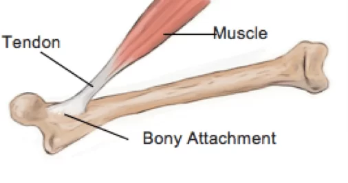
Collagen structure - Tendons are 85-95% Type I collagen organized in layers: tiny molecules → small fibers → bigger fibers → fiber bundles → whole tendon (Kjaer, 2004; Kannus, 2000). When you load a tendon, cells inside (tenocytes) sense the stretch and respond by making more collagen (especially Types I and III), creating stronger bonds between collagen fibers (cross-links), lining up fibers better along the pull direction, and making individual fibers thicker (Magnusson et al., 2010).
The signal pathway - Tendon cells have tiny sensors (integrin receptors) that detect when the tendon is being pulled or squeezed (Arnoczky et al., 2007). These sensors activate pathways inside the cell: FAK pathway increases gene activity, ERK/MAPK signaling makes collagen, TGF-β pathway remodels the structure, and COX-2 creates necessary inflammation for adaptation (Wang, 2006). No load = no signal = no adaptation.
Why Tendons Adapt So Slowly
Poor blood flow - Tendons have only 1-2% blood vessels compared to 15-20% in muscle (Kastelic et al., 1978). This means less oxygen and nutrients delivered, slower waste removal, fewer repair cells reaching the area, and collagen replaces at 1-2% per month versus 1-2% per day for muscle protein (Heinemeier et al., 2013).
Fiber reorganization - Resting collagen has a wavy "crimp" pattern (Franchi et al., 2007). Under load, waves straighten first (no strengthening yet), then fibers start bearing load. Months of training gradually reduce waviness, make fibers stiffer and better at storing energy, and improve bounce-back ability—taking 12-16 weeks minimum (Kongsgaard et al., 2009).
Why Different Training Methods Work
Isometric holds (holding still under tension, 30-45 seconds) - Create constant pressure that reduces abnormal blood vessels linked to pain (Cook & Purdam, 2009). Lower pain chemicals (glutamate and substance P) in the tendon and provide pain relief within 45 seconds through brain pain processing (Rio et al., 2015). Build strength without repeated stretching that might irritate inflammation—ideal for painful tendons.
Heavy slow resistance (lifting heavy weights slowly, 6-8 reps, 3-4 seconds up and down) - Loads over 70% maximum create enough stretch (4-8% deformation) to turn on collagen-making genes (Bohm et al., 2015). Slow movement maximizes time under tension, producing stiffer, stronger tendons with increased resistance to deformation (Arampatzis et al., 2007). This is the gold standard for long-term strengthening.
Eccentric training (focusing on lowering, 12-15 slow reps) - Lowering creates 20-50% more force than lifting (Roig et al., 2009). Creates tiny controlled damage triggering healing, disrupts abnormal pain-causing blood vessels, and lengthens muscle-tendon under load—a unique stimulus (Alfredson et al., 1998). Works best because tendons naturally experience highest forces when lowering.
Getting the Load Right
Too light (<30% max) - Not enough stretch to turn on collagen genes, maintains but doesn't strengthen, risks getting weaker over time (Magnusson et al., 2010).
Just right (70-85% max) - Creates 4-8% tendon strain, the sweet spot (Arampatzis et al., 2007). Activates cell sensors without damage, balances building versus breaking down (favors building), and activates enzymes creating cross-links (Kjaer et al., 2009).
Too heavy (>90% max or too much volume) - Exceeds repair ability, triggers too many breakdown enzymes (MMPs) causing net degradation, causes cumulative micro-damage, and leads to chronic tendon problems (Cook & Purdam, 2009).
Why Train 3-4 Times Weekly
Protein-making timeline - Collagen production peaks 24 hours after exercise, stays elevated 48-72 hours, returns to normal by 96 hours (Miller et al., 2005). Training every 48-72 hours catches the next session during elevated production—each builds on the last. But inflammatory chemicals (IL-1β, TNF-α) also peak at 24-48 hours, so you need rest days for inflammation to settle (Langberg et al., 2001).
Understanding Pain Levels
Why some pain is acceptable - Pain nerves cluster mostly in the outer sheath and bone attachment point, not the middle part (Ackermann et al., 2001). Middle loading can feel uncomfortable without causing damage. Pain at 2-3/10 that goes away within 24 hours = normal response, like muscle soreness (Silbernagel et al., 2007).
Why pain above 5/10 is dangerous - Indicates too much stretch or speed, may mean actual fiber tearing beyond healing capacity, triggers collagen-breakdown enzymes (MMP-13), and can cause bad blood vessel growth and nerve inflammation (Cook & Purdam, 2009).
The Stretching Issue
How stretching affects tendons - Tendons work like springs storing and releasing energy (Lichtwark & Wilson, 2006). Stretching before exercise makes tendons less stiff, reduces efficient force transfer, and can decrease force production 5-15% for 30-60 minutes after (Kubo et al., 2001). For injured tendons, excessive lengthening may worsen inflammation.
When stretching helps - Some mobility work maintains muscle-tendon length and prevents compensatory movements. Best done at different times than strength training (Magnusson et al., 2010).
The Adaptation Timeline
Weeks 0-4: Neural changes - Mainly nervous system improvements in muscle fiber recruitment with minimal tendon structure change (Kubo et al., 2002). Tendon cells start multiplying but remain vulnerable.
Weeks 4-12: Early structure changes - Collagen production increases (breakdown too), net positive building (Heinemeier et al., 2007). Fibers align better, stiffness gradually increases, but still relatively fragile.
Weeks 12-24+: Real strength - Cross-links increase, fibers get thicker, tendon size increases (visible on ultrasound), peak mechanical properties achieved (Kongsgaard et al., 2007). Biology cannot be rushed—jumping ahead means loading a tendon lacking the structure to handle it.
Why Wait for Jumping
The forces involved - Jumping creates forces 6-8 times body weight in milliseconds (Komi & Bosco, 1978). Requires stiffness developing only after months of training. Immature tendons lack enough cross-links for rapid stretch-release, risking complete tear or chronic pain (Kubo et al., 2002).
Safe progression - Isometric holds → heavy slow lifting → faster lifting → light jumping → full sport movements, over 4-6+ months minimum (Cook & Purdam, 2009).
Conclusion
Tendon strengthening is biological remodeling where cells respond to mechanical signals by making new, better-organized collagen. It's limited by poor blood supply and slow collagen turnover (30-60x slower than muscle). Success requires respecting these limits through appropriate loading, adequate recovery, and progressive increase over months. The difference between injury-resistant tendons and chronic pain is patience and evidence-based progression.
References
Ackermann, P. W., Li, J., Finn, A., Ahmed, M., & Kreicbergs, A. (2001). Autonomic innervation of tendons, ligaments and joint capsules. Journal of Orthopaedic Research, 19(3), 372-378.
Alfredson, H., Pietilä, T., Jonsson, P., & Lorentzon, R. (1998). Heavy-load eccentric calf muscle training for chronic Achilles tendinosis. American Journal of Sports Medicine, 26(3), 360-366.
Arampatzis, A., Karamanidis, K., Morey-Klapsing, G., De Monte, G., & Stafilidis, S. (2007). Mechanical properties of the triceps surae tendon and aponeurosis in relation to sport activity intensity. Journal of Biomechanics, 40(9), 1946-1952.
Arnoczky, S. P., Lavagnino, M., & Egerbacher, M. (2007). The mechanobiological aetiopathogenesis of tendinopathy. International Journal of Experimental Pathology, 88(4), 217-226.
Bohm, S., Mersmann, F., & Arampatzis, A. (2015). Human tendon adaptation in response to mechanical loading: A systematic review. Sports Medicine - Open, 1(1), 7.
Cook, J. L., & Purdam, C. R. (2009). Is tendon pathology a continuum? A pathology model to explain load-induced tendinopathy. British Journal of Sports Medicine, 43(6), 409-416.
Franchi, M., Trirè, A., Quaranta, M., Orsini, E., & Ottani, V. (2007). Collagen structure of tendon relates to function. TheScientificWorldJournal, 7, 404-420.
Heinemeier, K. M., Olesen, J. L., Schjerling, P., Haddad, F., Langberg, H., Baldwin, K. M., & Kjaer, M. (2007). Short-term strength training and myostatin and IGF-I isoforms in rat muscle and tendon. Journal of Applied Physiology, 102(2), 573-581.
Heinemeier, K. M., Schjerling, P., Heinemeier, J., Magnusson, S. P., & Kjaer, M. (2013). Lack of tissue renewal in human adult Achilles tendon. The FASEB Journal, 27(5), 2074-2079.
Kannus, P. (2000). Structure of the tendon connective tissue. Scandinavian Journal of Medicine & Science in Sports, 10(6), 312-320.
Kastelic, J., Galeski, A., & Baer, E. (1978). The multicomposite structure of tendon. Connective Tissue Research, 6(1), 11-23.
Kjaer, M. (2004). Role of extracellular matrix in adaptation of tendon and skeletal muscle to mechanical loading. Physiological Reviews, 84(2), 649-698.
Kjaer, M., Langberg, H., Heinemeier, K., Bayer, M. L., Hansen, M., Holm, L., Doessing, S., Kongsgaard, M., Krogsgaard, M. R., & Magnusson, S. P. (2009). From mechanical loading to collagen synthesis in human tendon. Scandinavian Journal of Medicine & Science in Sports, 19(4), 500-510.
Komi, P. V., & Bosco, C. (1978). Utilization of stored elastic energy in leg extensor muscles. Medicine and Science in Sports, 10(4), 261-265.
Kongsgaard, M., Reitelseder, S., Pedersen, T. G., Holm, L., Aagaard, P., Kjaer, M., & Magnusson, S. P. (2007). Region specific patellar tendon hypertrophy in humans following resistance training. Acta Physiologica, 191(2), 111-121.
Kongsgaard, M., Kovanen, V., Aagaard, P., Doessing, S., Hansen, P., Laursen, A. H., Kaldau, N. C., Kjaer, M., & Magnusson, S. P. (2009). Corticosteroid injections, eccentric decline squat training and heavy slow resistance training in patellar tendinopathy. Scandinavian Journal of Medicine & Science in Sports, 19(6), 790-802.
Kubo, K., Kanehisa, H., Kawakami, Y., & Fukunaga, T. (2001). Influence of static stretching on viscoelastic properties of human tendon structures. Journal of Applied Physiology, 90(2), 520-527.
Kubo, K., Kanehisa, H., & Fukunaga, T. (2002). Effects of resistance and stretching training on viscoelastic properties of human tendon structures. The Journal of Physiology, 538(1), 219-226.
Langberg, H., Rosendal, L., & Kjaer, M. (2001). Training-induced changes in peritendinous type I collagen turnover. The Journal of Physiology, 534(1), 297-302.
Lichtwark, G. A., & Wilson, A. M. (2006). Interactions between gastrocnemius muscle and Achilles tendon during incline, level and decline locomotion. Journal of Experimental Biology, 209(21), 4379-4388.
Magnusson, S. P., Langberg, H., & Kjaer, M. (2010). The pathogenesis of tendinopathy: Balancing the response to loading. Nature Reviews Rheumatology, 6(5), 262-268.
Miller, B. F., Olesen, J. L., Hansen, M., Døssing, S., Crameri, R. M., Welling, R. J., Langberg, H., Flyvbjerg, A., Kjaer, M., Babraj, J. A., Smith, K., & Rennie, M. J. (2005). Coordinated collagen and muscle protein synthesis in human patella tendon and quadriceps muscle. The Journal of Physiology, 567(3), 1021-1033.
Rio, E., Kidgell, D., Purdam, C., Gaida, J., Moseley, G. L., Pearce, A. J., & Cook, J. (2015). Isometric exercise induces analgesia and reduces inhibition in patellar tendinopathy. British Journal of Sports Medicine, 49(19), 1277-1283.
Roig, M., O'Brien, K., Kirk, G., Murray, R., McKinnon, P., Shadgan, B., & Reid, W. D. (2009). Effects of eccentric versus concentric resistance training on muscle strength and mass. British Journal of Sports Medicine, 43(8), 556-568.
Silbernagel, K. G., Thomeé, R., Thomeé, P., & Karlsson, J. (2007). Eccentric overload training for patients with chronic Achilles tendon pain. Scandinavian Journal of Medicine & Science in Sports, 11(4), 197-206.
Wang, J. H. (2006). Mechanobiology of tendon. Journal of Biomechanics, 39(9), 1563-1582.


